

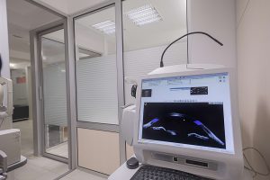
In plenty of up-to-date-equipped examination rooms, you will be welcomed by pleasant employees whose goal is to help you find solution for your health problem. Comprehensive ophthalmological examination involves observation of all ocular structures including anterior and posterior segment and the view of the problem entirely.
After initial ophthalmological examination, for detailed look into degree of defect or disorder and conclusion of the functional condition, specific modern technology diagnostic procedures are performed. Upon assessing the complete condition of the eye, eye specialist will explain the condition of the eye and recommend treatment.
VISUAL IMAGING:
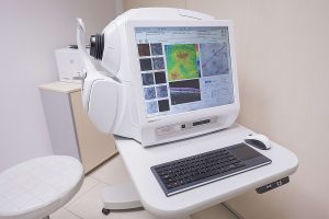
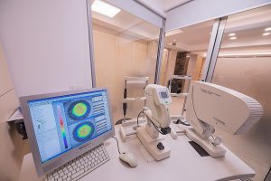
 OCT (OPTICAL COHERENCE TOMOGRAPHY)
Non-invasive, non-contact scanning procedure in which by using the light waves visual representation of sections through the eye are obtained, thickness and quality of cornea, appearance of anterior chamber, lens and specifically anterior chamber angle (greatest importance in narrow or closed angle glaucoma) which represents so called anterior OCT. Today it is unavoidable part of refractive procedures and also has important application in contactology in deciding for optimal contact lens.
Posterior OCT enables the sectional views of all retinal layers, particularly macula and the optic nerve. Today OCT represents a diagnostic procedure which is a must for a high- quality eye examination. It secures necessary guidelines for therapy of glaucoma and retinal diseases, especially macula (changes on macula lutea, edemas, macular degeneration, diabetic maculopathy).
Its significance is in localization of the impairment and monitoring of disease and therapy effects.
Combining it with fundus camera allows new possibilities of detection and locality of pathological event. It takes several minutes for the procedure.
 OCTA
OCTA
OCT Angio is the most recent non-invasive imaging technique, without contrast, without needle, for examining abnormal vasculatures through several sections and several layers (non-invasive angiology). The changes beneath the macular layer can be detected before occurrence of exudation and bleeding.
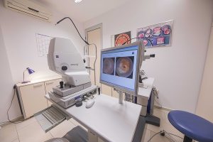 DIGITAL FUNDUS CAMERA AND ANGIOGRAPHY
They provide objective digital view of appearance and features of retina, macula lutea and optic nerve head. Using contrast intravenously, we find out the dynamics of circulation and complete view of blood vessels in retinal place, and we define the locus of occlusion (thrombosis), enlargement of blood vessel, bleeding etc.
FAF-FUNDUS AUTOFLUORESCENCA
Non-invasive diagnostic possibility with great importance for fast detection of photoreceptor damage and early metabolic changes at RPE level, which are base for genesis of degenerative, dystrophic macular damage, optic nerve head damage or retina entirely (atrophic macular degeneration, Stargardt, retinitis pigmentosa etc.).
ULTRASONIC DIAGNOSTICS
Standard diagnostic procedure for detection of deeper ocular structure changes, in vitreous body, retina and orbit (bleeding, retinal detachment, tumorous changes etc.), particularly in opaque conditions such as cataract or bleeding in anterior chamber which obstructs the view of the posterior structures.
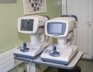
 IOL MASTER, STATE OF THE ART DEVICE – SWEPT SOURCE-
For multiple applications in ophthalmology. Precision measurement of the eye length (necessary for high myopia), thickness of the lens, anterior chamber depth, with fast detection of changes in deeper parts of the eye and macula lutea.
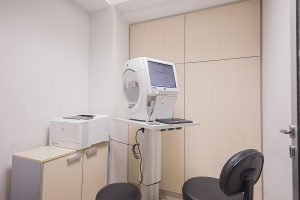
 PACHIMETRY AND COMPUTERIZED CORNEAL TOPOGRAPHY
Non-invasive imaging procedures obtaining the “map” of cornea with representation of all curvatures, strength of refraction and thickness in different points. Since cornea provides 2/3 of total refractive eye power (dioptric power of the eye), topography is of extraordinary importance for determining and assessment of corneal quality, early detection of irregularities and degenerative diseases (keratoconus, pellucid marginal degeneration), irregular astigmatism and changes occurring as a result of contact lens wearing etc. It is necessary for all refractive procedures.
 ENDOTHELIAL MICROSCOPY
Allows inspection of appearance, quality and quantity of endothelial cells, responsible for physiological function and regeneration of cornea along with assessment of the future bearing and decline ratio in numerous corneal dystrophies.
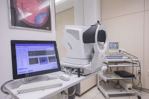
 COMPUTERIZED PERIMETRY
Investigation of the function of the optic nerve or macula – fovea. This diagnostic procedure objectively shows the level of functional decline in different diseases of the optic nerve head and macula.