ELECTRORETINOGRAPHY (ERG) and VISUAL EVOKED POTENTIAL (VEP) in eye practicenow is objective testing of the eye function, in children and adults.
The new way of thinking about modern visual electrophysiology.
Like EKG testing, it gives OBJECTIVE information about THE FUNCTION of retina and visual pathway by measuring the electrical responses of retinal cells upon the stimulation by light source. Various stimuli are usedto target different cellswithin the visual pathway to gain objective information about their function. We use VEP (visual evoked potential) and ERG (electroretinography), in several modalities and type of stimulation, depending on what we are testing for (pERG, ffERG, mfERG).


WHAT CAN WE TEST?
Retinal function in a variety of diseases:
-Glaucoma
– Optic neuritis
– CRVO (central retinal vein occlusion)
– Diabetic retinopathy
– Uveitis
– Toxic retinopathy
– Maculopathy (Macular degeneration, myopia, ERMA, cistoid, oedematous,.. )
– The retinal function could be tested as well in opaque media (cataract or haemorrhage in theeye), when is not possible to see the posterior pole in standard way.
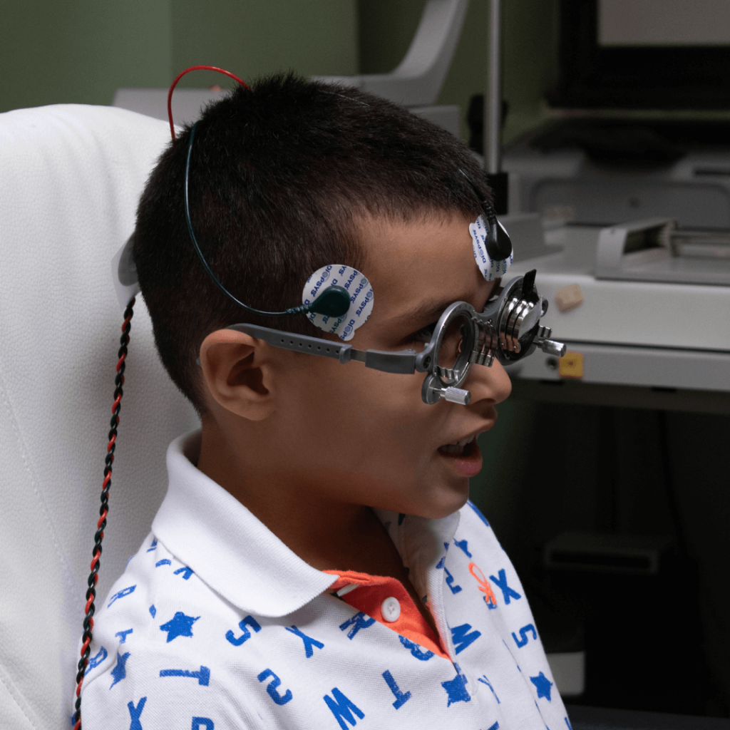
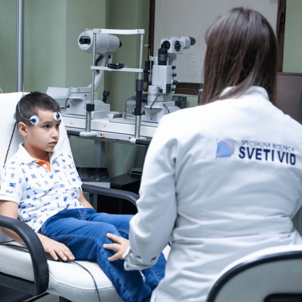
Easy to use in adults and in children, while seated comfortably and looking into flashing screen without stress and irritation of the eye. Only a few seconds testing, out-patient, without anaesthesia!
New modern visual electrophysiological testing gives us objective information about the damage of the eye function.
Thus is possible to detect even subtle changes in cell health, before clear clinical appearance of symptoms, or structural changes seen on OCT or digital fundus imaging, or defects depicted on computerized perimetry test.
OCT and fundus digital imaging are the brilliant way of seeing the retinal image, macula, nerve fiber layer and optic nerve head – structurally.
OCT is the best way of detecting ganglion cells damage, but, when they are already damaged (structural). Electrophysiology is the only way to detect the functional change, before structural damage!
CVP computerized visual field testing – perimetry, shows the defects in visual field, in glaucoma, optic neuritis, or macular impairments. It is one of the very important functional testing, but cannot show the early changing in visual pathway destruction.
Defects shown in visual field mean that already at least 30% of nerve fibers of the optic nerve are destroyed. This damage is irreversible. If discovered in that stage, we can treat against the further damage, but not recover already damaged nerve fiber layers.
With modern and comfortable visual electrophysiology, we can now even predict the future damages and more precisely decide WHAT AND WHEN TO TREAT.
BY TRACKING THE TREATMENT EFFICACY, in follow-ups, the evaluation of tests over time, will show when to go on, or stop with the treatment (e.g. drops, laser, anti-VEGF,…) or when to plan the surgery.
UNLIKE THE OLD PLATFORM OF ELECTROPHYSIOLOGY used in neuro-ophthalmology, for decades ago, within almost two hours testing, invasive electrode placement directly on conjunctiva, touching the cornea, in the eye, with a lot of irritation and tears, with anaesthesia, subjective, in reading the results, THIS NEW MODERN CONCEPT OF ERG and VEP is:
-objective,
-repeatable
-easy to use
-comfortable for children and adults
-non-invasive,
-without touch of the eye – electrodes are placed beneath the lower lids, not in the eye
-without anesthesia
-lasting only few minutes,
-fast and reliable answer of retinal and visual function.